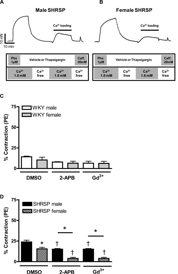
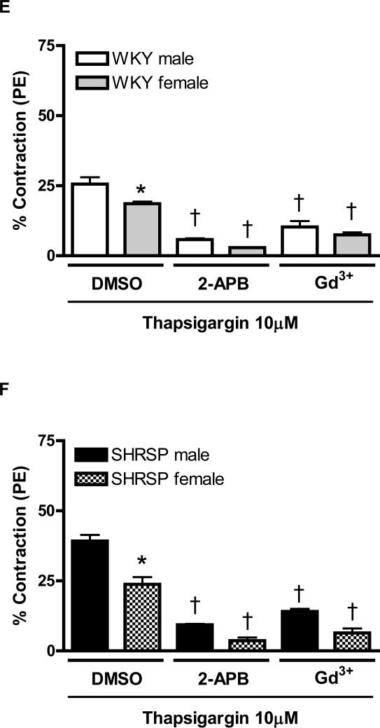
Figure 1. Increased contraction upon Ca2+ loading period, in aortas from male compared to female SHRSP, is normalized after Orai1/CRAC channel inhibition.
The tracings illustrate the protocol used to evaluate force in response to Ca2+ influx after depletion of intracellular Ca2+ stores (loading period) in aortas from (A) male and (B) female SHRSP rats. Aortic rings were contracted with PE (1μM) and then, washed in Ca2+ free -EGTA buffer, in the presence or absence of thapsigargin (10μM). After 20 minutes, 1.6mM Ca2+ buffer was added and aortic rings were incubated for an additional 15 minutes with or without thapsigargin. Aortic rings were washed with Ca2+ free-EGTA buffer for 2 minutes and then stimulated with caffeine (20mM). This protocol was performed in aortas from (C, E) male (white bars) and female (grey bars) WKY rats, or in aortas from (D, F) male (black bars) or female (hatched bars) SHRSP rats, in the (C, D) absence or in the (E, F) presence of thapsigargin. Note that the contractions were significantly inhibited after CRAC channel blockade with 2-APB and Gd3+. Values are expressed as mean ±SEM. * P< 0.05 vs. respective male; † P< 0.05 versus respective DMSO.


