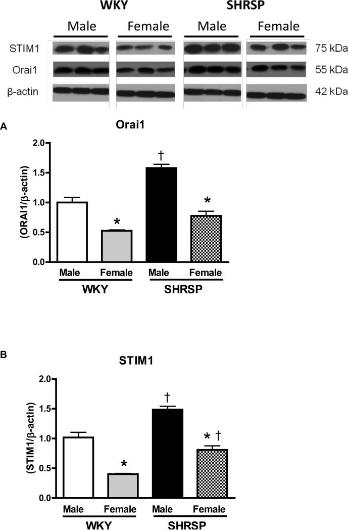
Figures 4. Aorta from male SHRSP display increased Orai1 and STIM1 protein levels, compared to arteries from female SHRSP.
Top, representative western blot images and bottom, corresponding bar graphs demonstrating that (A) Orai1 and (B) STIM1 protein are augmented in male SHRSP aortas, compared to aortas from male WKY or female SHRSP. Densitometric analyses were performed and values were normalized to β-actin protein expression. * P< 0.05 vs. respective control; ‡ P<0.05 vs. respective male.

