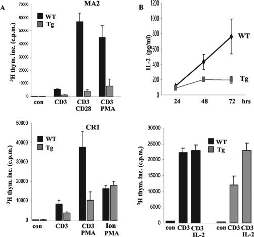
FIG. 3.
SinΔC expression inhibits T-cell proliferation and IL-2 production. (A) T cells (105) purified from the spleens of 6- to 8-week-old normal and SinΔC animals (CR1 and MA2) were stimulated with plate-bound CD3 and CD28 antibodies and PMA and ionomycin as indicated and as described in Materials and Methods. Proliferation was calculated by [3H]thymidine incorporation (3H thym. inc.) as the mean ± SD of radioactive counts in triplicate wells. Shown are data representative of at least three experiments with two different transgenic lines (MA2, top panel; CR1, bottom panel). (B) Five microliters of the supernatants was removed from stimulated cells at the indicated times and analyzed by enzyme-linked immunosorbent assay for the presence of secreted IL-2 (top graph). Purified T cells were stimulated with plate-bound anti-CD3 antibody (0.05 μg/ml) in the presence or absence of IL-2 (20 ng/ml). Proliferation was determined by [3H]thymidine incorporation (bottom graph). Tg, transgenic; WT, wild type.

