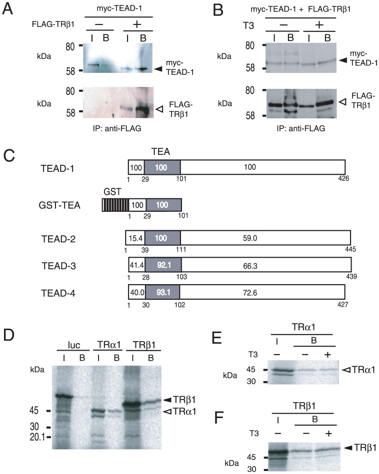
Figure 7. TRs directly interact with TEAD-1.
(A) Co-immunoprecipitation of TRβ1 with TEAD-1. The expression plasmids for 6myc-TEAD-1 (pcDNA3-6myc-TEAD-1) were transfected into CV-1 cells with or without FLAG-TRβ1. Whole-cell extracts were immunoprecipitated with the anti-FLAG M2 affinity gel and analyzed by Western blotting with the anti-myc antibody. I, input; B, bound; IP, immunoprecipitation. The numbers on the left side of each panel indicate molecular mass markers (kDa). Solid and open arrowheads indicate 6myc-TEAD-1 (upper panel) and FLAG-TRβ1 (lower panel), respectively. (B) The interaction of TRβ1 with TEAD-1 was T3-independent. In the presence or absence of 1 µM T3, co-immunoprecipitation of TRβ1 with TEAD-1 was performed under the same conditions as in (A). (C) Schematic representations of TEAD-1, GST-TEA, and TEAD-2, 3, and 4. TEA, the TEA domain. The numbers within and under the box represent amino acid homology (%) and codon numbers, respectively. (D) Both TRα1 and TRβ1 interact directly with the TEA domain of TEAD-1. A GST pull-down assay was performed using GST-TEA and 35S-labeled in vitro translated TRα1, TRβ1, or firefly luciferase (luc). (E) and (F) The interaction of TEAD-1 with TRα1 (E) or TRβ1 (F) is T3-independent. A GST pull-down assay was performed in the presence or absence of 1 µM T3 under the same conditions as (D). The numbers on the left side of each panel indicate molecular mass markers (kDa). Solid and open arrowheads indicate TRα1 and TRβ1, respectively. I, input; B, bound.

