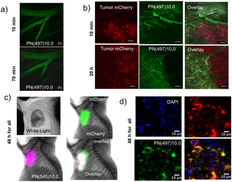
Figure 5. Fluorescent imaging of three pharmacokinetic phases of PN’s with diameters of 10 nm.
A) Vascular phase, two photon microscopy of brain vasculature of normal mice. Mice underwent a craniotomy and implantation of a transparent window. Vessel intensity drops due vascular escape, but there is no interstitial fluorescence in the brain due to the blood brain barrier. Scale marker = 50 microns. B) Intravital confocal microscopy of the vascular and interstitial phases of an mCherry expressing HT-29 xenograft. During the vascular phase (10 min post-injection), vessels are imaged, without interstitial fluorescence. During the interstitial phase (20 h post-injection), interstitial fluorescence is prominent. Scale marker = 20 microns. C) Surface fluorescence/X-ray imaging of the tumor retention phase of PN(545)10.0. Shown are the HT-29/mCherry tumor with the skin removed as a white light image, mCherry tumor fluorescence (green), PN(545)10.0 fluorescence (purple) and the green/purple over lay (white). D) Confocal microscopy of the tumor retention phase of PN(497)10.0. Shown are a sectioned HT-29 mCherry expressing tumor with nuclei stained blue (DAPI), mCherry tumor cells (red), PN(497)10.0 (green) and a green/red overlay (yellow).

