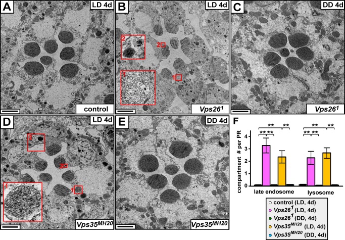
Figure 3. Late endosomes and lysosomes are expanded in Vps26 and Vps35 mutant PRs.
(A) TEM of the PRs of a control (iso) fly kept in LD for 4 d show normal morphology with intact rhabdomeres and cell bodies. Scale bar, 1.5 µm. (B) Upon 4 d in LD, Vps261 mutant PRs exhibit highly aberrant morphological features: note the expansion of late endosomes (multivesicular structures, inset 1) and lysosomes (condensed structures, inset 2). In addition, electron dense particles are increased, and the morphology of some rhabdomeres is defective compared to control in (A). Scale bar, 1.5 µm. (C) Upon 4 d in DD, Vps261 mutant PRs exhibit similar morphology as controls. Scale bar, 1.5 µm. (D) Upon 4 d in LD, Vps35MH20 mutant PRs show increased late endosomes (inset 1) and lysosomes (inset 2) similar to Vps261 mutants. Scale bar, 1.5 µm. (E) Upon 4 d in DD, the morphology of Vps35MH20 mutant PRs remains similar to that of the control. Scale bar, 1.5 µm. (F) Quantification of the number of late endosomes and lysosomes in PRs at day 4 in LD or DD. Ten PRs were quantified for each genotype. Student's t test; error bars represent SEM; ** p<0.01.

