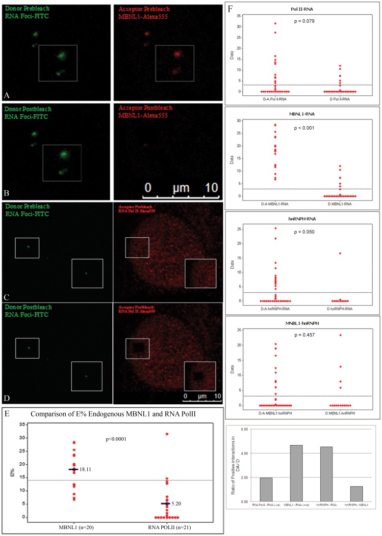
Figure 1. Evidence of intracellular interaction between endogenous MBNL1, and hnRNPH with RNA foci using AP-FRET.
RNA foci in DM1fibroblasts were detected by RNA-FISH (green) in combination with immunofluorescence for either endogenous MBNL1 or RNA Pol II (red). FITC-Alexa555 was used as the FRET pair. Representative donor and acceptor pre-bleach (A and C) and post-bleach (B and D) images are shown for MBNL1-RNA foci or RNA Pol II foci AP-FRET. Dequenched signal from the donor (FITC) was seen after photobleaching the acceptor demonstrating interaction of MBNL1 with RNA foci. (E) Comparison of E% distribution for FRET assays of RNA foci and MBNL1 interactions in DM1 cells (n = 20) and for RNA foci and RNA Pol II interactions (n = 21). (F) Comparison of E% distribution in FRET assays for interactions of RNA Pol II, MBNL1, and hnRNPH with RNA foci, and for interactions of hnRNPH and MBNL1. A normalized ratio of the number of positive ROIs in Donor/Acceptor (DA) to positives in Donor only (D) FRET assays underscores that both hnRNPH and MBNL1 interact with the RNA foci. Line on graphs represents the positive FRET threshold level.

