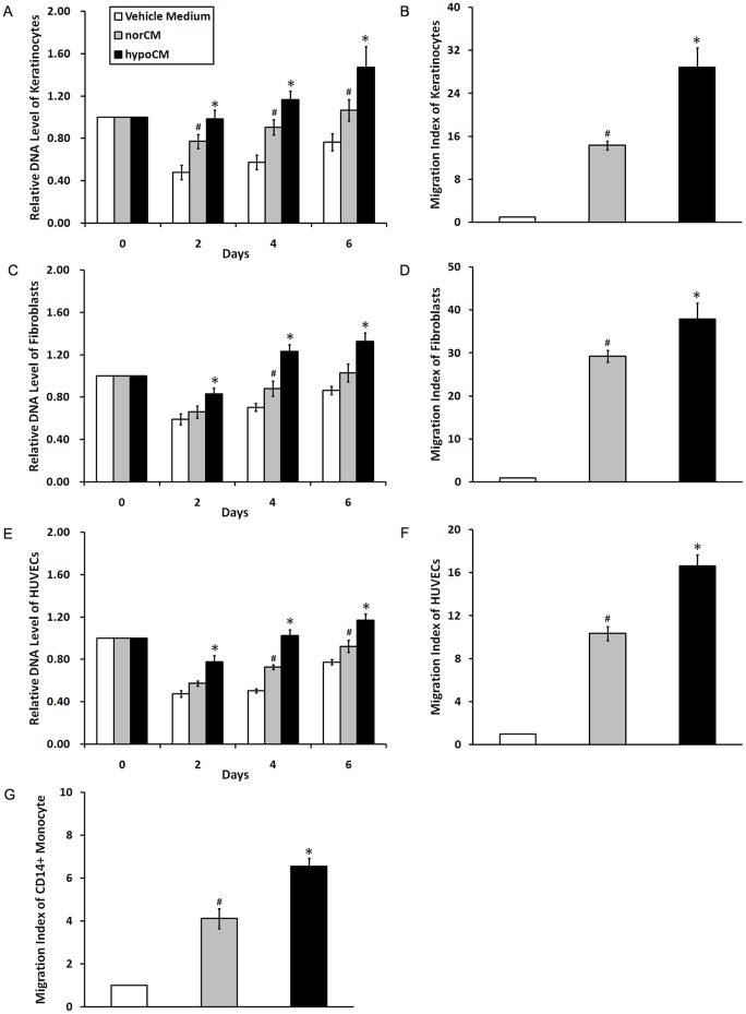
Figure 3. Effects of BM-MSC-derived conditioned medium samples on paracrine cell proliferation and migration.
Equal numbers of keratinocytes, fibroblasts and HUVECs were incubated with vehicle control medium, norCM or hypoCM. Cell proliferation was evaluated at indicated time points (A, C and E). Data are given as the means±the SEM; *p< 0.05 compared with the vehicle control or the norCM group. #p< 0.05 the vehicle control compared with the norCM group. Equal numbers of keratinocytes, fibroblasts, HUVECs and CD14+ monocytes were added to the upper chambers of 24-well transwell plates, with the indicated medium added to the lower chambers (n = 4 wells per treatment). Cells that migrated to the bottom of the filter were stained and evaluated (B, D, F and G). Data are given as the means± the SEM;*p< 0.05 compared with the vehicle control or the norCM group. #p< 0.05 the vehicle control compared with the norCM group.

