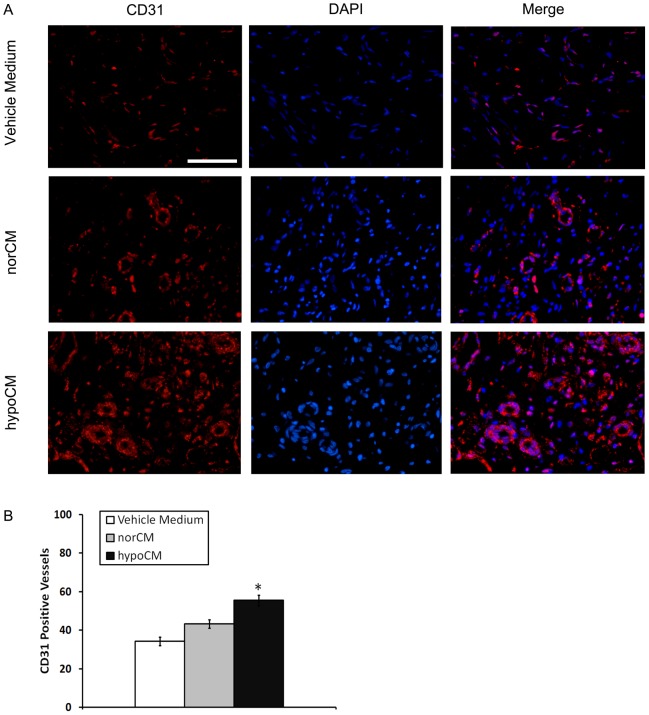
Figure 7. Angiogenesis in the murine skin after wounding.
Wound sections were evaluated on day 11 by staining with anti-CD31 antibody. Representative CD31+ vessels are shown (A). The extent of vascularization was determined by assessing the number of CD31+ vessels in each of 4 randomly chosen high-power fields within the injury site (B). Scale bar, 100 µm for images in (A) (400×). Results are given as the means ± the SEM; *p< 0.05 compared with the vehicle control or the norCM group.

