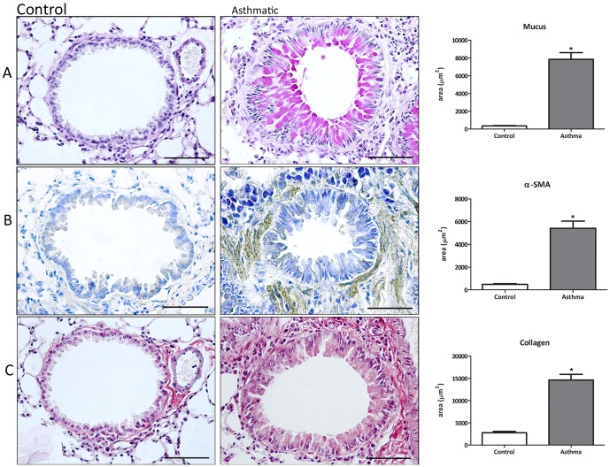
Figure 3. Fibrosis markers in the asthmatic lung.
Lung sections were taken from animals with asthma (6 challenges), stained with PAS for mucus (A) or prepared for immunohistochemical and morphometric analysis of α-SMA (B) and collagen type I (C). Data are representative of 5 animals in each of 3 independent experiments. Bars = 50 mm. *p<0.05 comparing with control group.

