FIG. 1.
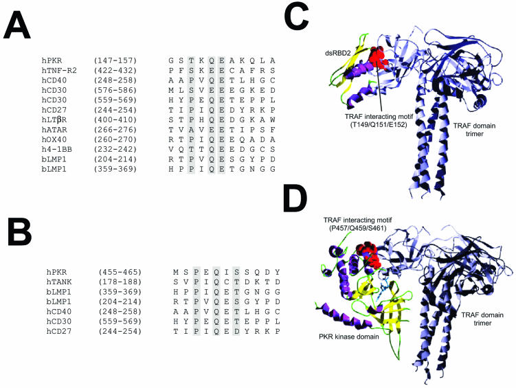
Model predicting TRAF-PKR interaction. (A) Sequence alignment of the putative TRAF-interacting motif present in the dsRBD2 subdomain of PKR with those from different TRAF binding domains. (B) Sequence alignment of the putative TRAF-interacting motif present in the kinase domain of PKR with those from different TRAF binding domains. (C) Putative model of the interaction between dsRDB2 and TRAF. A ribbon plot of the structure obtained through docking procedures for the interaction between the TRAF-interacting motif (T149/Q151/E152; red spheres) of the dsRDB2 subdomain of PKR and TRAF. TRAF trimer secondary-structure elements are depicted in grey. The dsRBD amino-terminal domain of PKR is shown as a ribbon plot (α helices, magenta; β strands, yellow). (D) Model of the PKR (kinase domain)-TRAF interaction. A docking model of the putative interaction between the TRAF-interacting motif (P457/Q459/S46; red spheres) of the kinase domain of PKR and the TRAF domain. TRAF trimer secondary-structure elements are depicted in grey. In the kinase domain of PKR, α helices are in magenta and β strands are in yellow. A molecule of ATP has been included solely to indicate the position of the putative ATP binding pocket.

