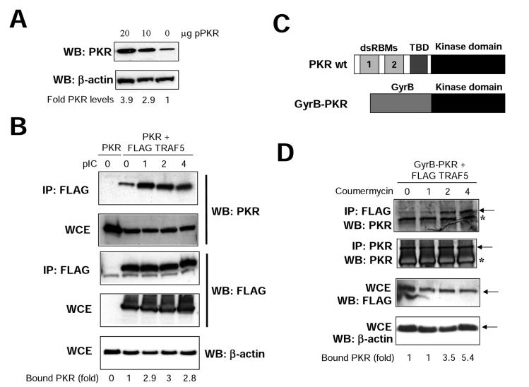
FIG. 5.
TRAF-PKR interaction is inducible. (A) 293T cells growing in 10-cm-diameter dishes were transfected with 10 or 20 μg of a plasmid coding for PKR, and at 48 h after transfection, cell extracts were collected and analyzed by immunoblotting (Western blotting [WB]) with antiserum to PKR or actin. Signals were quantitated with NIH Image software. (B) 293T cells were transfected with 20 μg of a plasmid coding for PKR (lane 0) or with 10 μg each of plasmids encoding for PKR and FLAG-TRAF5, as indicated. At 48 h after transfection, cells were treated with 10 μg of pIC per ml. At the noted times, cell extracts were collected, immunoprecipitated (IP) with anti-FLAG serum, and thoroughly washed and immunocomplexes or whole-cell extracts (WCE) were analyzed by SDS-PAGE and subjected to immunoblotting with antiserum to PKR, FLAG, or actin. Signals were quantitated with NIH Image software. (C) Schematic representation of PKR and the GyrB-PKR chimera. The different domains of PKR are shown (dsRBMs, the third basic domain [TBD], and the kinase domain). The chimera consists of the N-terminal 220 aa of E. coli GyrB fused to the kinase domain (aa 258 to 551) of human PKR. (D) 293T cells were transfected with 10 μg each of plasmids encoding Gyr-PKR and FLAG-TRAF5 as indicated. At 48 h after transfection, cells were treated with coumermycin. At the indicated times posttreatment, cell extracts were collected, immunoprecipitated with anti-FLAG or anti-PKR serum, and thoroughly washed and immunocomplexes or whole-cell extracts (WCE) were analyzed by SDS-PAGE and subjected to immunoblotting with antiserum to PKR, FLAG, or actin. Asterisks denote IgGs, and arrows indicate the specific proteins. Signals were quantitated with NIH Image software.

