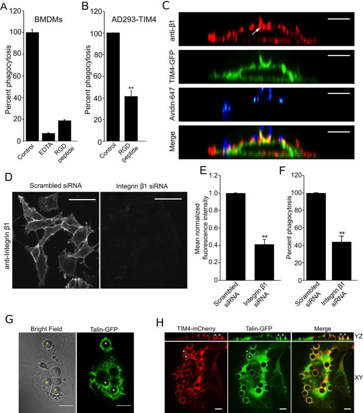
FIGURE 4:
Integrin β1 is required for TIM4-dependent phagocytosis. (A) Wild-type BMDMs were treated with 10 mM EDTA or 500 μg/ml RGD peptide, and uptake of PtdSer-bearing beads was assessed. Data were normalized to untreated control and are means ± SEM of three independent experiments. (B) Effect of RGD peptide on phagocytosis of PtdSer-bearing beads by AD293 TIM4 transfectants. Data were normalized to untreated control and are means ± SEM of three independent experiments. (C) Distribution of integrin β1, detected by immunostaining, in the phagocytic cup of AD293 TIM4 transfectants. The representative micrographs show YZ reconstruction of images obtained by spinning disk confocal microscopy. The white arrow indicates the localization integrin β1 to the TIM4-GFP–positive phagocytic cup. Incompletely engulfed portions of the target were labeled with fluorescent avidin and are shown in blue. Bar, 6.8 μm. (D) Representative fluorescence micrographs showing immunofluorescence staining for endogenous integrin β1 after treatment of AD293 TIM4 transfectants with either scrambled siRNA (left) or integrin β1-specific siRNA (right). Bar, 17.1 μm. (E) Quantitation of the effect of integrin β1-specific and scrambled siRNA on β1 protein expression, from experiments like that in D, quantifying the mean fluorescence intensity of endogenous integrin β1 after immunostaining. Data were normalized to scrambled siRNA–treated cells and are the mean ± SEM of three independent experiments. (F) Effect of integrin β1-specific and scrambled siRNA on TIM4-mediated phagocytosis of PtdSer beads in AD293 TIM4 transfectants. Data were normalized to scrambled siRNA–treated cells and are the mean ± SEM of three independent experiments. (G) Fluorescence micrographs showing confocal images of wild-type BMDMs expressing talin-GFP engulfing PtdSer beads. Asterisks indicate accumulation of talin-GFP at nascent phagosomes. Scale bar, 10 μm. (H) Fluorescence micrographs showing confocal images of AD293 cells expressing TIM4-mCherry and talin-GFP engulfing PtdSer beads. Asterisks, two representative phagocytic cups that can be seen in the YZ reconstruction on top, showing TIM4-mCherry and talin-GFP accumulation. Scale bar, 6.8 μm. **p ≤ 0.01.

