Figure 2.

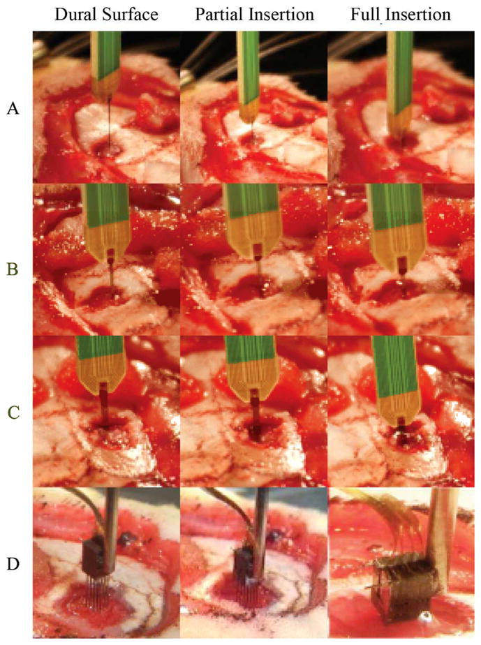
Still-frame images of probe insertions into rat brain. a–c) Shanks 1, 2, 4 shank probes respectively, all inserted with minimal dimpling d) 3D probe inserted with increased dimpling

Still-frame images of probe insertions into rat brain. a–c) Shanks 1, 2, 4 shank probes respectively, all inserted with minimal dimpling d) 3D probe inserted with increased dimpling