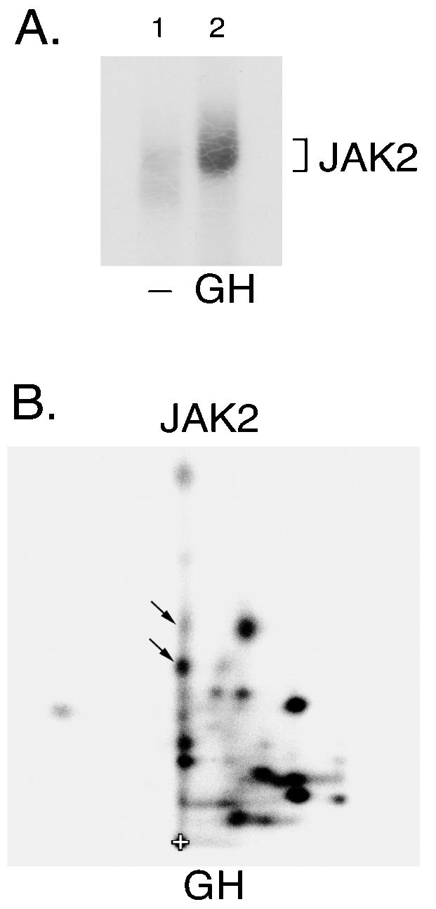
FIG. 3.

JAK2 activated in response to GH autophosphorylates tyrosine 813. (A) 3T3-F442A cells were incubated in the absence (lane 1) or presence of 30 ng of GH/ml (lane 2) for 15 min. The cells were lysed and JAK2 immunoprecipitated using anti-JAK2. JAK2 was immobilized and incubated in the presence of [γ-32P]ATP at 30°C for 30 min. The JAK2 was resolved by SDS-PAGE, transferred to nitrocellulose and visualized by autoradiography. (B) 32P-labeled JAK2 was cut from the nitrocellulose and subjected to 2-D peptide mapping with the thin-layer electrophoresis step performed at pH 1.9 (5). The origin (+), and spots whose migration are similar to spots identified in Fig. 1A are indicated.
