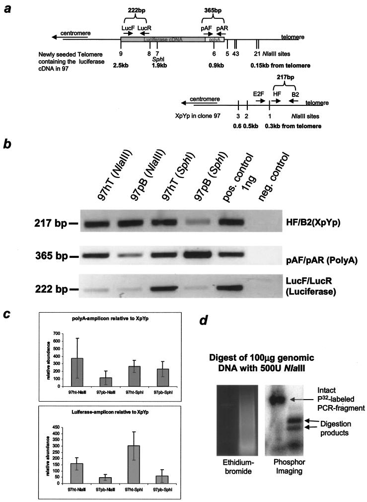
FIG.4.
PCR analysis of the X-region at a newly seeded telomere in 97hT and 97pB. (a) Map of the first nine NlaIII sites and one SphI site and their distance from the telomere, which also has been confirmed with sequence analysis of a newly seeded telomere and the Xp/Yp telomere in 97hT and 97pB. (b) Representative PCR amplification from telomere preparations after NlaIII or SphI digestion using primer pairs that are located in the subtelomeric region. (c) Semiquantitative analysis using spot densitometry (AlphaImager software) from three independent telomere preparations. The results of each amplification have been normalized to a standardized positive control (1 ng of genomic DNA of 97), and the averages from eight different PCRs have been normalized to total telomere input (XpYp PCR). (d) Control showing the complete digestion by NlaIII of a radioactively labeled PCR fragment mixed with genomic DNA (before telomere preparation), shown on an ethidium bromide-containing gel, and the subsequent PhosphorImager analysis.

