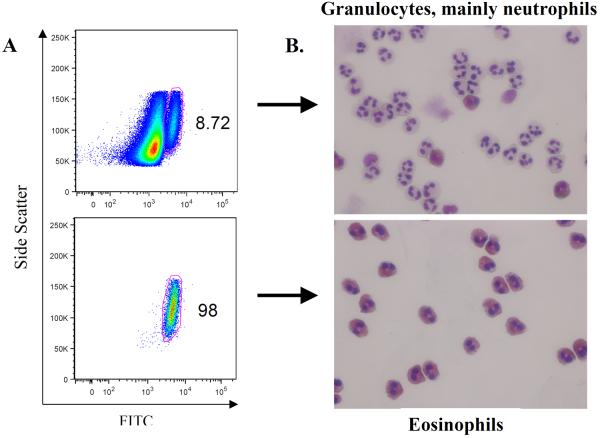
Figure 1. Purity of isolated human eosinophils.
(A) Eosinophils were identified by their forward scatter, side scatter, and autofluorescent properties on flow cytometry of unstained human granulocyte and isolated eosinophil preparations. While neutrophils and isolated eosinophils have similar forward and side scatter characteristics, eosinophils have higher autofluorescence, as captured by the FITC channel. (B) Cytospin preparations of granulocytes and eosinophils underwent modified Wright-Giemsa staining. Neutrophils can be distinguished from the characteristic pink, granular cytoplasm of the eosinophils. The upper panel for both (A) & (B) represents cells derived from the initial granulocyte isolation which consists of primarily neutrophils. The lower panel for both (A) & (B) represents the granulocyte cell population after eosinophil enrichment.

