
Figure 2.
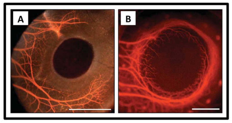
Corneal nerves during development. A) Formation of the chick pericorneal nerve ring at embryonic day 6 (E6). Fluorescence image with brightfield overlay. B) Completion of the chick pericorneal nerve ring and subsequent penetration of corneal nerves into the stroma at E8. Fluorescence image only. Scale bar for A and B: 1 mm. Courtesy of Dr. James K. Kubilus, Department of Anatomy and Cell Biology, Tufts University School of Medicine, Boston, MA. 113