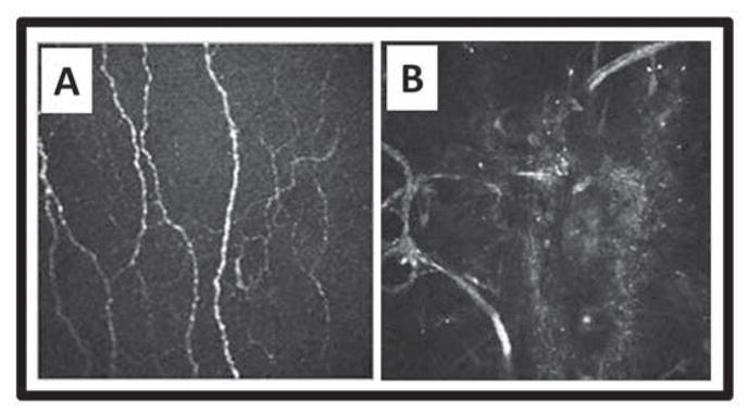
Figure 6.

Subbasal nerves before and after LASIK. Heidelberg Retina Tomograph II with Rostock Corneal Module optical coherence tomography showing subbasal corneal nerves A) before LASIK, and B) regenerated nerve loops in the flap area 4 years postoperatively. Courtesy of Heidelberg Engineering, Inc. and Dr. E.M. Messmer, Munich, Germany.
