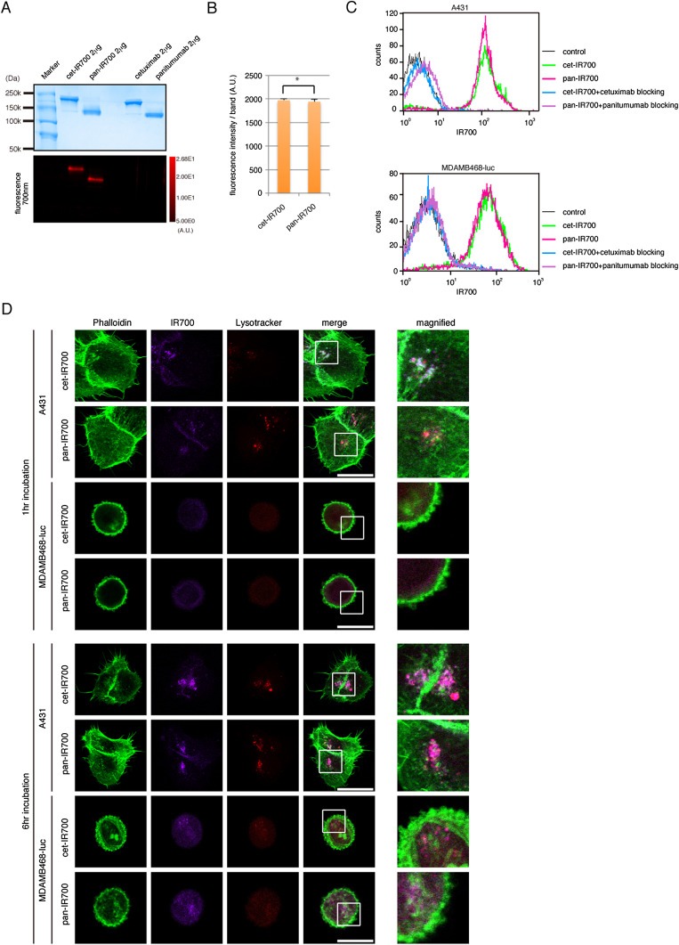
Figure 1.

Quality control of cet‐IR700 and pan‐IR700 synthesis. (A) Validation of cet‐IR700 and pan‐IR700 by SDS‐PAGE (upper: colloidal blue staining, lower: fluorescence). Diluted commercial cetuximab and panitumumab were used as controls. (B) Quantification of fluorescence intensity between cet‐IR700 and pan‐IR700 (*ns). (C) Specific binding function to EGFR by flow cytometry showing similar binding. (D) A431 and MDAMB468‐luc cells were incubated with cet‐IR700 or pan‐IR700 for indicated times and fixed. Immunostaining was performed with Lyso Tracker (red: lysosome detection) and phalloidin (green: actin detection, especially membrane). Both cet‐IR700 and pan‐IR700 were internalized into cells in a time dependent manner. Bar = 25 μm.
