
Figure 3.
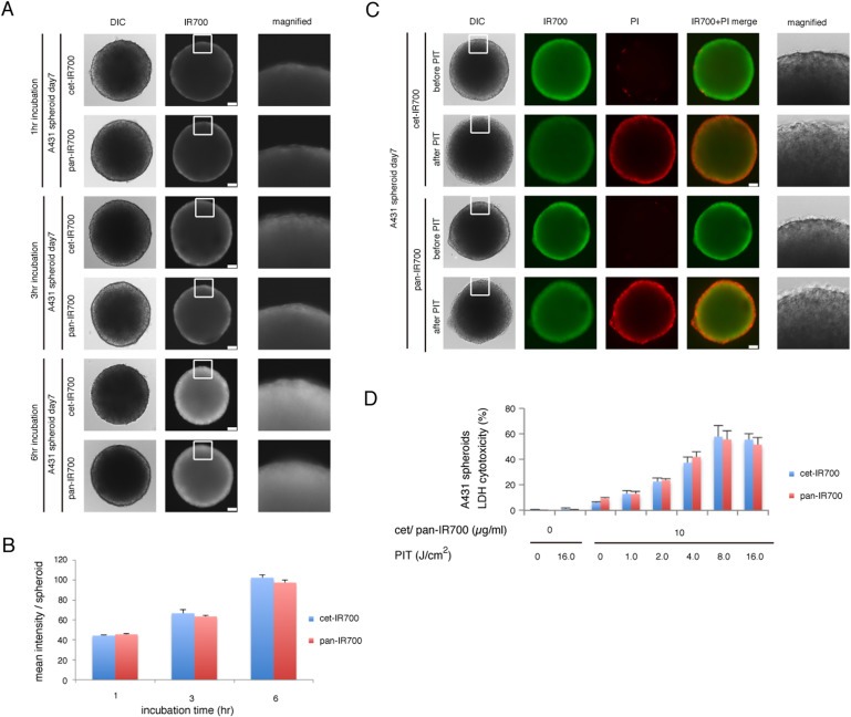
Permeation of cet‐IR700 and pan‐IR700 into A431 3D spheroids and the effect on PIT. (A) Permeation of cet‐IR700 or pan‐IR700 into A431 3D spheroids increased over time. Bar = 100 μm. (B) Quantification of the intensity of IR700 showed no significant difference between cet‐IR700 and pan‐IR700 (n = 10) at any point. (C) A431 3D spheroids were treated with PIT after exposure to either cet‐IR700 or pan‐IR700 for 6 h and observed by microscopy (before and after irradiation of NIR light). Necrotic cell damage was observed after PIT. Bar = 100 μm. (D) LDH cytotoxicity assay (for the spheroids exposed with cet‐IR700 and pan‐IR700 for 6 h) showed increasing cell death with increases in light dose. There was no significant difference between cet‐IR700 and pan‐IR700 in efficacy at any point.