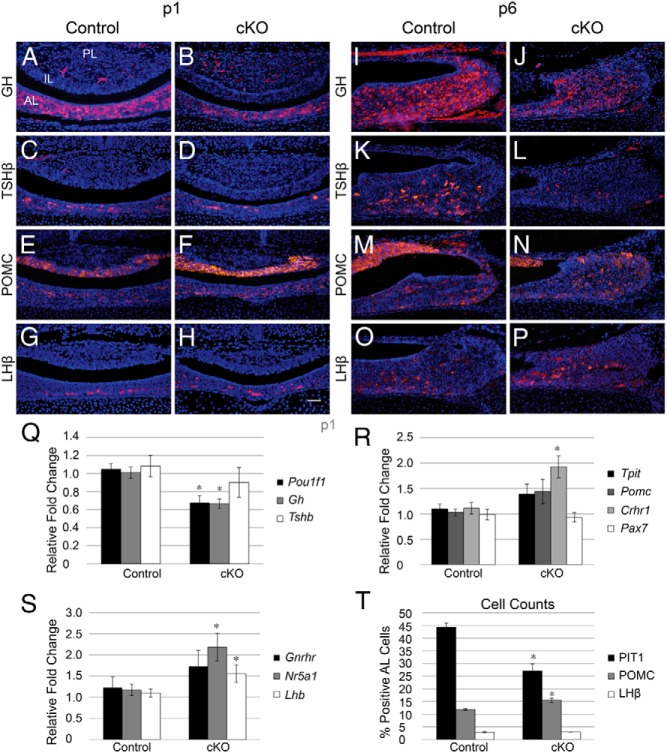
Figure 6.
Conditional loss of Notch2 results in decreased expression of Pit1 lineage hormones and increased expression of POMC. GH-immunopositive cells are found scattered throughout the AL of control mice (A) and appear to be reduced in pituitaries of cKO mice (B) at p1. TSHβ is localized to the AL of control (C) and cKO (D) mice. POMC-immunoreactive cells are found in the AL and the IL of control (E) pituitaries and appear slightly increased in the AL of cKO mice (F). LHβ expression is confined to the AL and appears similar between control (G) and cKO (H) mice. At p6, GH is present in the AL of control mice (I) and appears reduced in cKO mice (J). Similarly, TSHβ is strongly reduced in cKO mice (L) compared with those in control mice (K). POMC is present in the IL and AL of control mice (M) and appears slightly increased in the AL of cKO mice (N). LHβ appears similar between control (O) and cKO (P) pituitaries. qRT-PCR analysis at p1 reveals a significant decrease in Pou1f1 (Pit1) and Gh mRNA, but no change in Tshb mRNA between control and cKO mice (Q). In addition, p1 mRNA levels of Tpit and Pomc show a trend toward an increase in cKO mice, whereas Crhr1 is significantly increased and Pax7 is unchanged (R). mRNA levels of Nr5a1 and Lhb are significantly increased in pituitaries, whereas Gnrhr is trending toward an increase at p1 (S). The percentages of Pit1-, POMC-, and LHβ-positive cells in the AL were quantified in control and cKO mice, and the percentage of Pit1-positive cells was found to be deceased in cKO mice, whereas the percentage of POMC-immunoreactive cells is increased (T). *, P < .05. Magnification, ×200. Bars, 50 μm. n = 5 to 7 (histology), n = 4 to 7 (qRT-PCR), and n = 3 to 5 (cell counts).

