
Figure 7.
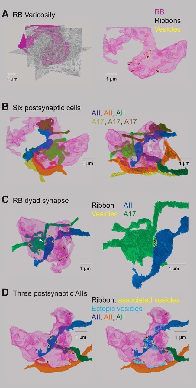
Detailed reconstruction of an RB terminal varicosity. A, Left, An RB terminal varicosity (magenta) superimposed on SBEM sections. Right, The same varicosity contains several synaptic ribbons (green) and their associated vesicles (cyan). Not all of the ribbons in this varicosity are illustrated. B, Three AII amacrine cells (bluish) and three A17 amacrine cells (brownish) are postsynaptic to the ribbons illustrated in A. Right and left show the same cells from two different perspectives. C, A dyad synapse. Left, A single ribbon (arrowhead) is presynaptic to an AII and to an A17. The dyad is shown in more detail at right. D, Left, Three AIIs are postsynaptic to the ribbons illustrated. Right, Vesicles near the plasma membrane but unassociated with ribbons are presynaptic to the AIIs.