Figure 3.

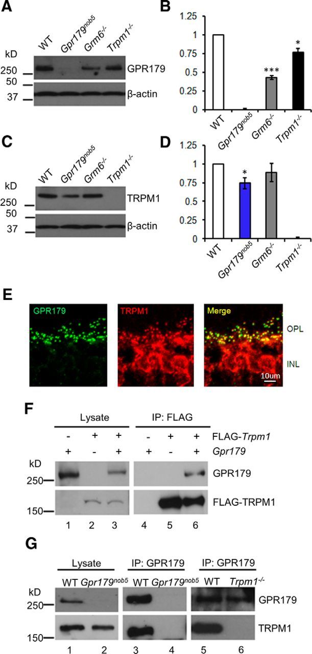
GPR179 and TRPM1 proteins interact. Western blots of total retinal lysates probed with antibodies to (A) GPR179 and (C) TRPM1 (top). Each blot was reprobed with antibodies to β-actin (bottom) to determine total protein and for use as an internal standard. Band intensities were analyzed and quantified with NIH ImageJ software and normalized to β-actin expression level in the same sample. The histograms (B, D) plot the mean expression (±SEM) from four experiments on independent retina samples. B, GPR179 expression was lower in Grm6−/− and Trpm1−/− retinas compared with WT. D, TRPM1 expression was similar to WT in Grm6−/− and in Gpr179nob5 retinas. Statistical analyses (t test) were performed before normalization. Error bars represent SE; *p < 0.05, ***p < 0.001. E, GPR179 and TRPM1 colocalize on the dendritic tips of DBCs. Representative confocal images of cross sections of the OPL and inner nuclear layer (INL) of WT retina reacted with antibodies to TRPM1 (green) and GPR179 (red). The merged image shows that TRPM1 and GPR179 expression colocalize at the dendritic tips of DBCs. Scale bar, 5 μm. F, Western blot of lysate from HEK293T cells transfected with plasmids expressing GPR179 (lane 1), FLAG-TRPM1 (lane 2), or both (lane 3) and probed with antibodies to GPR179 (top row) and FLAG (bottom row). The presence of a specific expression construct is indicated by “+” above the lane on the blot. Lysates from HEK293T samples (lanes 1–3) were immunoprecipitated with antibodies to GPR179 and the precipitates analyzed by Western blotting (lanes 4–6), using antibodies to GPR179 (top row) or TRPM1 (bottom row). These data show that TRPM1 is coimmunoprecipitated with GPR179 (lane 6). G, Western blot of retinal lysates from WT (lane 1) and Gpr179nob5 (lane 2) probed with antibodies for presence of GPR179 (top row) or TRPM1 (bottom row). Western blots of proteins coimmunoprecipitated with antibodies to GPR179 from WT (lanes 3,5), Gpr179nob5 (lane 4), and Trpm1−/− (lane 6) probed for GPR179 (top row) or TRPM1 (bottom row). IP with GPR179 antibody from retinal lysates of Gpr179Nob5 and Trpm1−/− mice served as controls for nonspecific binding. These data were representative of at least three independent experiments using independent retinal samples. Data show that GPR179 and TRPM1 coimmunoprecipitate (lanes 3,5).
