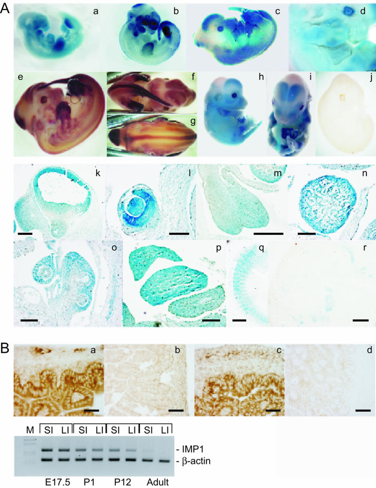
FIG.2.
Expression of Imp1 during development and in adult tissues. (A) β-Galactosidase activity (a, c, d, h, i, and k to q) and Imp1 mRNA expression revealed by whole-mount in situ hybridization with an antisense Imp1 riboprobe (b, e, f, g, and j) was examined in E10.5 (a and b), E12.5 (c to g), E14.5 (h and i)m and E17.5 (r) embryos. No Imp1 mRNA expression was observed in Imp1−/− E12.5 embryos (j). E12.5 embryos were sectioned, and β-galactosidase activity was detected in snout and forebrain (k), eye (l), tongue (m), heart (n), lung (o), liver (p), and somites (q). Moreover IMP1 protein expression was detected in kidneys from E17.5 embryos (r). Bars: k, 350 μm; l, 125 μm; m, 250 μm; n, 250 μm; o, 140 μm; p, 250 μm; q, 250 μm; r, 120 μm. (B) Immunohistochemistry was performed to detect IMP1 in the small intestine (a) and large intestine (c) from E17.5 embryos. No staining was observed in adult large intestine (d). As a control IMP1-specific antibodies were preadsorbed with the peptide that was used for the immunizations (b). Bars: a to d, 60 μm. In the lower panel, the expression of Imp1 mRNA was examined in small and large intestines from E17.5, P1, P12, and adult mice by multiplex RT-PCR. Specific primers for Imp1 and β-actin, resulting in 237-bp and 157-bp PCR fragments, respectively, were used. The products were separated by agarose gel electrophoresis and visualized by ethidium bromide staining. SI, small intestine; LI, large intestine.

