FIG. 7.
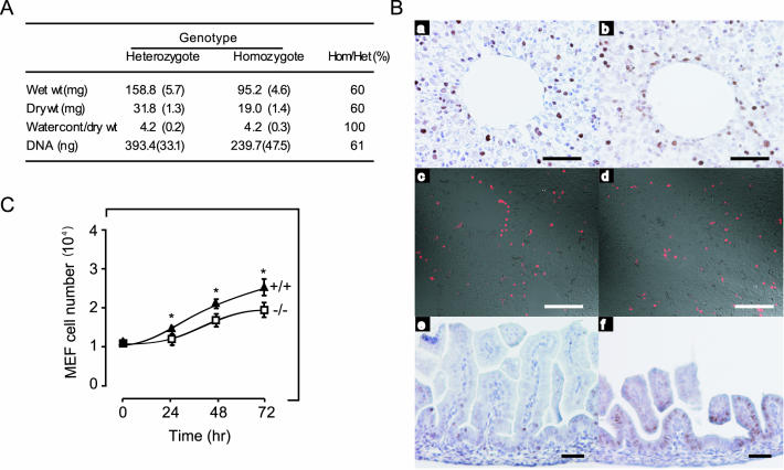
Reduced cellular proliferation in organs and MEFs from Imp1−/− mice. (A) The wet weight, dry weight, and DNA content of the kidney from 3- to 4-month-old Imp1+/− (n = 3) and Imp1−/− (n = 3) mice were analyzed as described in Materials and Methods. (B) Liver (a to d) and small intestine (e and f) sections from wild-type and Imp1−/− E17.5 embryos, respectively, were examined for proliferation by PCNA staining (a, b, e, and f) or for apoptosis by TUNEL staining (c and d). (C) Growth of MEFs from wild-type (▴) and Imp1−/− (□) E13.5 embryos. Cells were plated in 30-mm plates and counted for 4 consecutive days. Each value is an average of three independent experiments with duplicate plates. Growth of Imp1−/− MEF cells was significantly (P < 0.05) reduced after 24, 48, and 72 h. Scale bars: 200 μm (A and B), 300 μm (Card D), and 60 μm (E and F).

