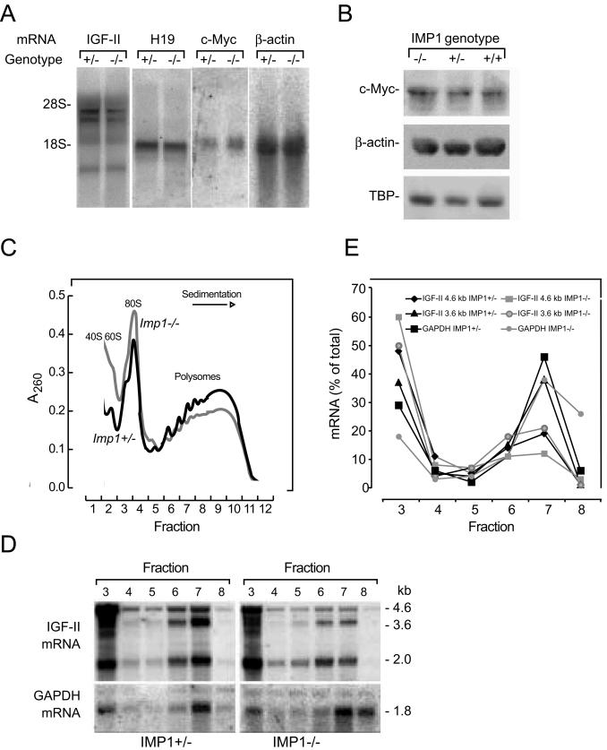
FIG. 8.
Expression of IMP1 target genes in E12.5 embryos. (A) Northern blot analysis of Igf2, H19, c-myc, and β-actin mRNAs in Imp1+/− and Imp1−/− E12.5 embryos. Total RNA (10 μg) was hybridized to cDNAs encoding Igf2, H19, c-myc, and β-actin. (B) Western blot analysis of c-Myc and β-actin in wild-type, Imp1+/−, and Imp1−/− whole E12.5 embryo lysates. Extracts were separated in SDS-10% polyacrylamide gels, transferred to Hybond-P membranes, and probed with antibodies specific for c-Myc and β-actin. As a loading control, the membranes were probed with an anti-TBP antibody. (C and D) Isolation of polysomes from E12.5 embryos. Cytoplasmic lysates from Imp1+/− and Imp1−/− embryos were separated in a 20 to 47% sucrose gradient. Panel C shows the A260 sedimentation profile of ribosomal subunits and polyribosomes, and panel D shows the Northern blot analysis of Igf2 mRNA in the fractions. The position of the major Igf2 leader 3 (4.6-kb) and leader 4 (3.6-kb) mRNA species derived from mouse promoters P2 and P3, respectively, are indicated. (E) Relative levels of Igf2 4.6-kb and 3.6-kb mRNAs in polysomes and monosomes, respectively. Data are represented as a percentage of total photon-stimulated luminescence for each transcript.

