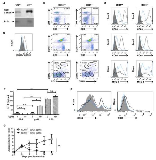
Figure 1. Selective loss of CD91 in CD11c+ cells renders mice unresponsive to gp96.
CD91flox/flox mice were mated with CD11c+-Cre recombinase mice and backcrossed. (A) BMDCs were sorted for CD11c to 96% purity and analyzed by immunoblotting for CD91 β-chain or actin as a loading control. (B) Lymph node cells from CD91−/− (blue) or CD91+/+ littermates (black) were stained with anti-CD91 antibody, gated on CD11c+ and analyzed by flow cytometry. Filled grey is secondary antibody alone. (C) Lymph node cells from CD91−/− or CD91+/+ littermates were phenotyped for markers indicated. (D) BMDCs from CD91−/− or CD91+/+ mice were pulsed on day 6 with LPS for 24 hours (black line) or left un-pulsed (blue line). Cells were analyzed for expression of CD86, CD40 and MHC II. Filled grey is secondary antibody alone. (E) BMDCs from CD91−/− or CD91+/+ mice were pulsed with 100μg gp96 or 1EU LPS for 24 hours or with PBS. IL-1β was measured by ELISA. (F) BMDCs from CD91−/− (blue line) or CD91+/+ (black line) mice were pulsed on day 6 with 100μg gp96 for 24 hours or left un-pulsed (filled grey). Cells were analyzed for expression of CD86 and CD40. (G) CD91−/− or CD91+/+ mice were immunized with 1 μg E.G7 tumor-derived gp96 twice, one week apart, followed by E.G7 tumor challenge one week later. Tumor growth was monitored. * P< 0.05, ** P< 0.01, n.s. not significant. Error bars indicate s.e.m. Experiments were independently performed twice with 2 (A-F) or 3-5 (G) mice per group.

