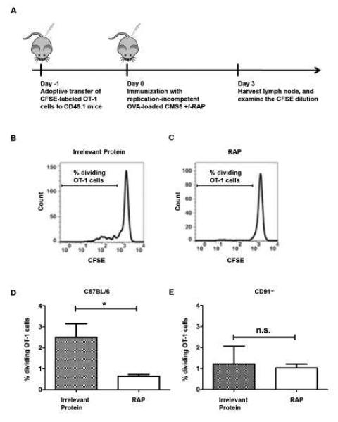
Figure 5. Inhibition of antigen cross-presentation by RAP reduces T cell proliferation in vivo.
The effect of RAP on T cell proliferation in vivo was examined. (A) CD45.1+ C57BL/6 mice were adoptively transferred with CFSE-labeled CD45.2+ OT-1 cells (1.5×10⁶ per mouse), one day before immunization with mitomycin-C treated OVA-loaded CMS5 with or without RAP expression. After 3 days, the draining inguinal and axillary lymph nodes were harvested and stained for CD45.2 and CD8 expression to differentiate adoptively transferred OT-1 cells from endogenous responses. (B and C) Histograms from the flow cytometry analysis of gated CD45.2+CD8+ OT-1 cells from representative mice of each group. (D) The percentages of dividing OT-1 cells were compared in mice immunized with OVA-loaded CMS5 cells, expressing RAP or control protein. (E) CD8+OT-1 proliferation in CD91−/− mice was measured on day 3 following immunization with mitomycin-C treated OVA-loaded CMS5 with or without RAP expression. The percentages of dividing OT-1 cells are shown. * P < 0.05, n.s. not significant. Experiments were independently performed twice with 3-5 mice per group. Error bars indicated s.e.m.

