Fig. 2.
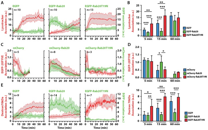
The association of Rab20 with phagosomes delays phagosome maturation. (A) The kinetics of LTR acquisition by phagosomes in macrophages expressing EGFP, EGFP–Rab20 or EGFP–Rab20T19N. RAW264.7 macrophages were transfected with EGFP, EGFP–Rab20 or EGFP–Rab20T19N and incubated with LTR for 30 min. Cells were subsequently incubated with 3-µm IgG-coated beads and analyzed by using live-cell imaging. (B) Data points from A at 5, 15 and 60 min were pooled. (C) The kinetics of the association of PI3P with phagosomes in macrophages expressing mCherry, mCherry–Rab20 or mCherry–Rab20T19N. RAW264.7 macrophages were co-transfected with EGFP–2xFYVE and mCherry, mCherry–Rab20 or mCherry–Rab20T19N, incubated with 3-µm IgG-coated beads and analyzed by using live-cell imaging. EGFP–2xFYVE and mCherry fluorescence were pseudo-colored to red and green, respectively. The intensity of both EGFP and mCherry fluorescence was normalized to the fluorescence intensity of the cytoplasm. (D) Data points from C at 5, 15 and 60 min were pooled. (E) The kinetics of the delivery of 70-kDa dextran to phagosomes in macrophages expressing EGFP, EGFP–Rab20 and EGFP–Rab20T19N. RAW264.7 macrophages were transfected with EGFP, EGFP–Rab20 or EGFP–Rab20T19N and preloaded with Texas-Red-conjugated 70-kDa dextran (50 µg/ml) for 2 h, followed by washing and chasing for 16 h. Afterwards, cells were incubated with 3-µm IgG-coated beads and analyzed by using live-cell imaging. (F) The data points from E at 5, 15 and 60 min were pooled. n, the number of phagosomes analyzed in at least three different experiments. All data show the mean±s.d. of the intensity at 5, 15 and 60 min post-internalization from three independent experiments. *P≤0.05, **P≤0.01, ***P≤0.001 (Student's two-tailed unpaired t-test).

