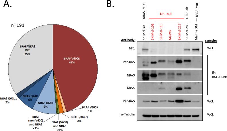
Figure 1.
NF1-null melanoma cell lines express high levels of activated RAS. A) BRAF and NRAS status of the melanoma cell line panel (n=191). B) Activated RAS protein (RAS-GTP) was quantitated in select melanoma cell lines via immunoprecipitation with the RAS-binding domain of RAF (RAF-1 RBD) followed by immunoblot using pan-RAS and isoform selective NRAS and KRAS antibodies. Expression of NF1, total RAS (pan-RAS) and actinin (as a loading control) were measured by immunoblot from whole cell lysate (WCL). Alt = alteration, mut = mutant, WCL = whole cell lysate.

