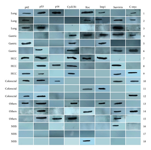
Figure 1.

Miniarray of multiple TAAs with representative cancer sera using western blot analysis. Lanes 1–3 are three representative lung cancer sera; lanes 4–6 are three representative gastric cancer sera; lanes 7–9 are three representative hepatocellular carcinoma (HCC) sera; lanes 10–12 are three representative colorectal cancer sera; lanes 13–15 are three representative other cancers sera, showing different antibody profiles with the eight TAAs; and lanes 16–18 are three representative normal human sera (NHS), showing positive reactivity to Koc and survivin but not with other TAAs.
