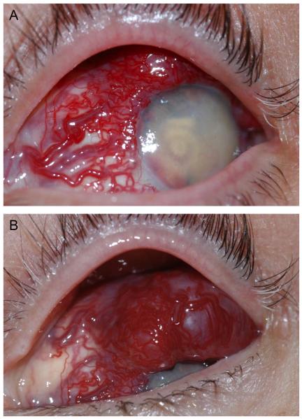
Figure 1.
External photos of the highly vascularized lesion. A. Dilated tortuous vessels involving the nasal, superior, and temporal conjunctiva feed a highly vascularized subconjunctival lesion. The cornea is translucent; there is florid neovascularization with a layered hyphema in the anterior chamber. B. Views of the superior conjunctiva reveal that the lesion overrides the corneal limbus superiorly.

