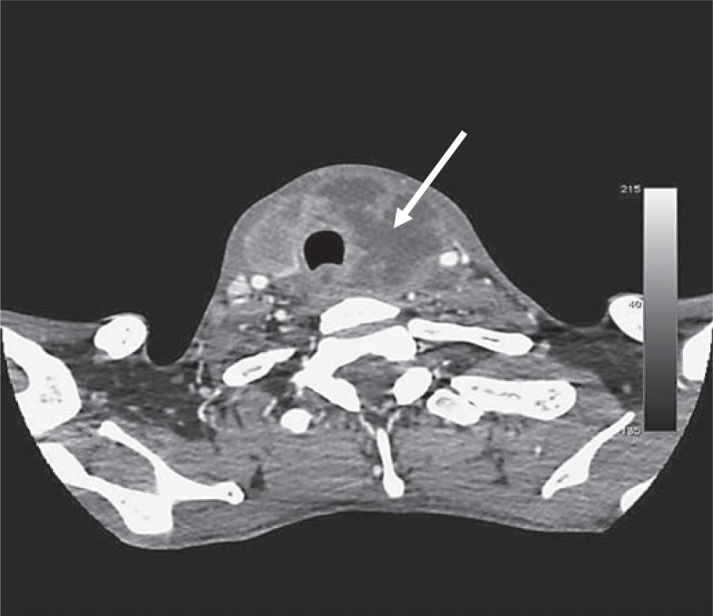
Fig. 1.
Computed tomography scan of the patient's neck showing a large rim-enhancing collection (45 × 45 × 52 mm) in the left lobe and isthmus of the thyroid (arrow). The abscess extended around the trachea, compressing the esophagus. The right lobe showed an inhomogeneous pattern. There were significant paratracheal and jugular lymph nodes.

