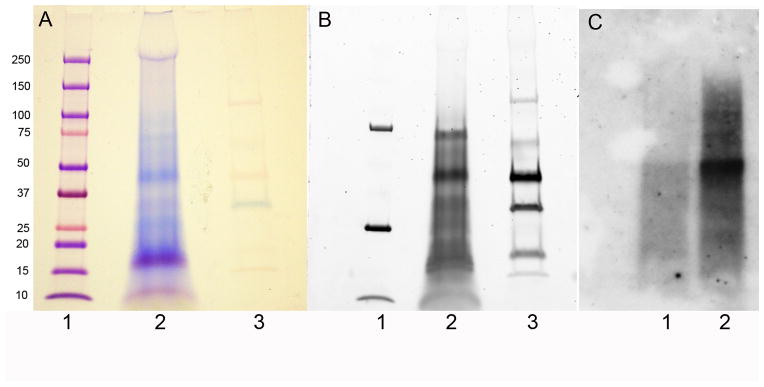
Figure 2. SDS-PAGE of proteins present in the 0.6 N HCl extract of Lytechinus variegatus tooth.

In A the gel was stained with Stains All. In B the same gel was stained with ProQ Diamond phosphoprotein staining Kit prior to staining with Stains all. In C the gel was transferred to nitrocellulose and over laid with Ca45. Lanes A1 and B1 are molecular weight standards. Lanes A2 and B2 are 0.6 N HCl extracts and lanes A3 and B3 are phosphorylated standards. In C lane 1 is the 6M Guanidine Hydrochloride extract and lane 2 is 0.6 N HCl extract
