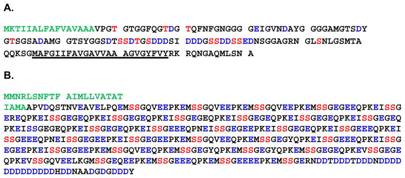
Figure 3. The amino acid composition of Lytechinus variegatus P16 and Phosphodontin.
A is the sequence of P16. The signal peptide is highlighted in green. The potential phosphorylation sites are highlighted in red and the acidic amino acids are highlighted in blue. The transmembrane domain is underlined. B is the sequence of Phosphodontin: The signal peptide is highlighted in green. The Ser-Ser pairs which are casein kinase 2 substrates are highlighted in red, while the acidic amino acids are highlighted in blue.

