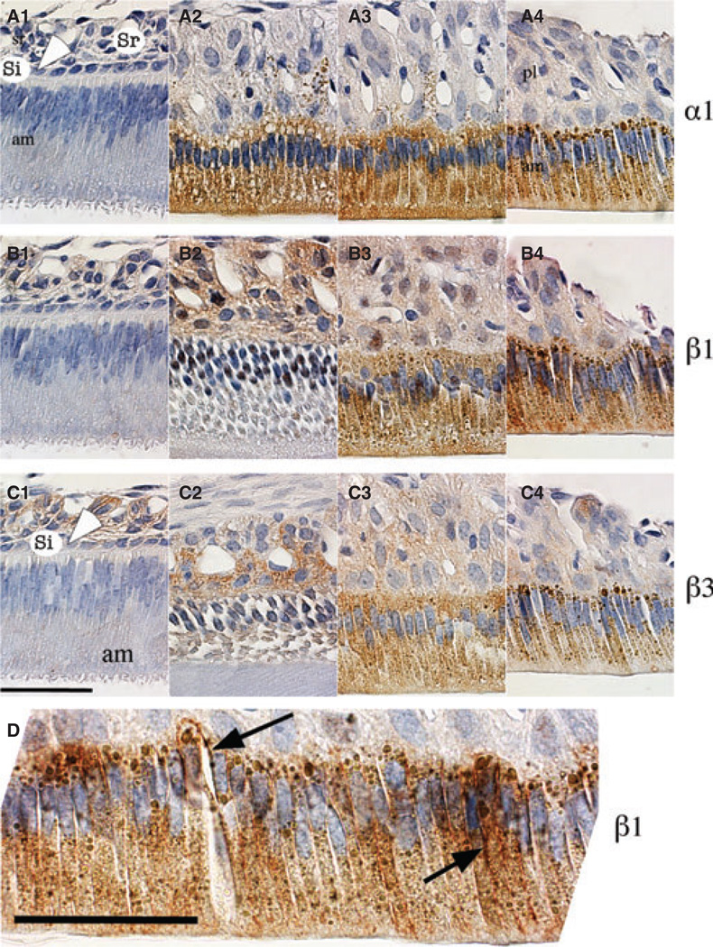
Fig. 3.
Immunolocalization of α1, β1, and β3 subunits in enamel organ of rat incisors. Sagittal sections of maxillary incisors from 4-wk-old rats were stained with antibodies against subunit α1 (panels A1–A4), β1 (panels B1–B4), and β3 (panels C1–C4) and were counterstained with haematoxylin. Differential localization is shown at secretory (panels A1, B1, and C1), early-maturation (panels A2–3, B2–3, and C2–3) and late-maturation (panels A4, B4, and C4) stages of enamel organ development. Enamel organ cells, ameloblasts (am), stratum intermedium (si, and white arrowhead), stellate reticulum (sr), and papillary layer cells (pl) are labelled. (D) Magnified image of late-maturation-stage ameloblasts illustrating the basolateral (black arrow) and cytoplasm expression, possibly associated with the endoplasmic reticulum, of the β1 subunit. Scale bars = 50 µm.

