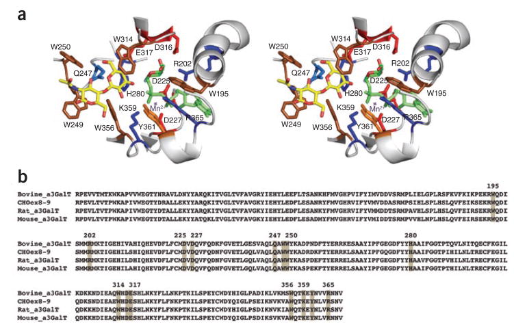
Figure 1.
Structural complex of CHO Ggta1 exons 8–9 gene product with UDP-2F-Gal donor and LacNAc acceptor substrate. (a) Shown in stereo view is the cartoon representation of the enzyme active site with the side chains of the key residues labeled using the single amino acid code and the numbering based on the bovine α3GalT crystal structure (PDB: 1G93). The UDP-2F-Gal is shown in stick representation in green and the type II LacNAc acceptor substrate is shown in stick representation in yellow. The location of the Mn2+ cation (in purple) is obtained from the bovine α3GalT crystal structure template used to construct the homology model. (b) Sequence alignment of the various α3GalT with the highly conserved critical active site residues highlighted in gray.

