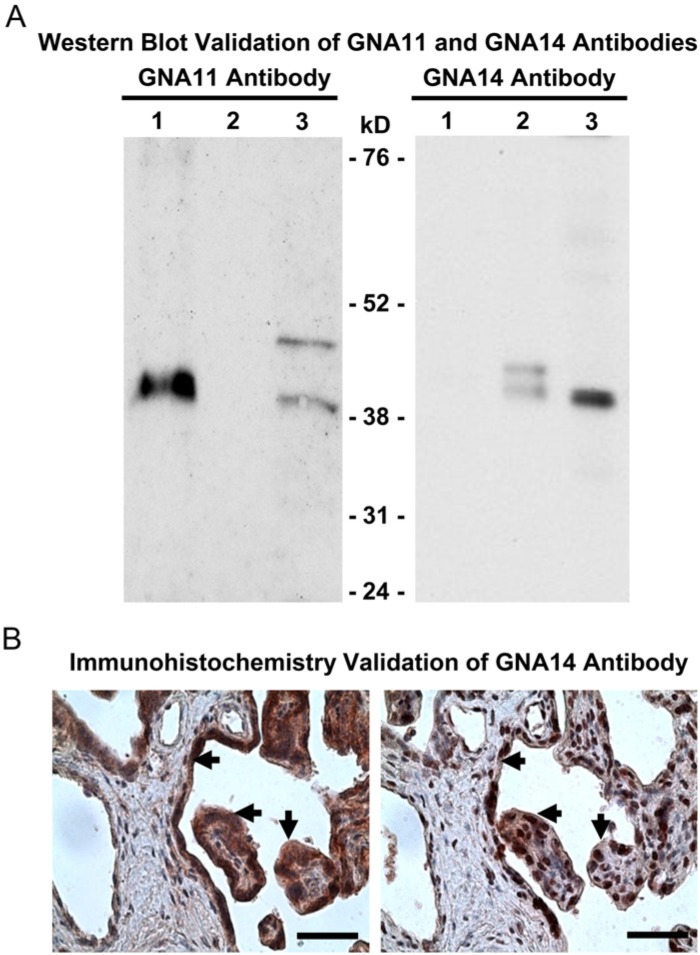
Figure 1.
Validation of GNA11 and GNA14 antibodies. (A) Western blot validation of GNA11 and GNA14 antibodies. Lane 1: GNA11-overexpressing 293T cells (10 µg protein); Lane 2: GNA14-overexpressing 293T cells (10 µg protein); Lane 3: HUVECs (20 µg protein). (B) Immunohistochemistry validation of GNA14 antibody in human placentas (n=4) from severe preeclamptic (sPE) pregnancies. After counterstaining with hematoxylin, the adjacent tissue sections were probed with the GNA14 antibody alone (4 μg/ml, left panel) or the GNA14 antibody pre-immunoneutralized with its peptide immunogen (right panel). Representative images are shown. Arrows: syncytiotrophoblasts. Bar, 50 µm.

