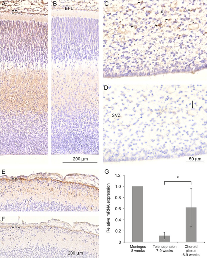
Figure 1.
YKL-40 protein distribution in the neocortex of a human fetus at the 12th week post-conception (wpc) (CRL: 75 mm) (A-B), and in the occipital cortex of a 21st wpc fetus (CRL: 200 mm) (C-F). Sections shown in (A), (C) and (E) were stained with the monoclonal antibody (MAb) 201.F9 whereas those in (B), (D) and (F) were stained using the MAb 201.F9 preabsorbed with purified human YKL-40 protein. Note the lack of or a markedly diminished reactivity of the sections stained with the preabsorbed YKL-40 antibody. The end feet layer (EFL) is barely visible in (B) and (F), and the presumed astroglial progenitors (arrow) in the subventricular zone (SVZ) in (C) is no longer positive following preabsorption (D). Note the reactivity associated with the blood vessel in (C) (arrow) and the lack of reactivity in (D) (arrow). (G) Quantitative real-time RT-PCR analysis of YKL-40 mRNA expression in the human developing forebrain from tissue samples dissected from two human embryos (aged 6 weeks and 5 days post-conception and 7 wpc) at the 7th and 8th wpc and three human fetuses (aged 8 wpc, 8 weeks and 3 days post-conception, and 9 wpc) at the 9th and 10th wpc. Relative mRNA expression is shown (meninges=1). Only one meninges sample was available for analysis. A significant difference between average expression values obtained in the telencephalon (n=4) and the choroid plexus (n=3) was observed (indicated by *, Students t-test, p=0.029). Error bars represent one standard deviation. Scale bar: A–B: 200 μm, C–D: 50 μm, E–F: 200 μm. Same magnification in (A-B), (C-D) and (E-F).

