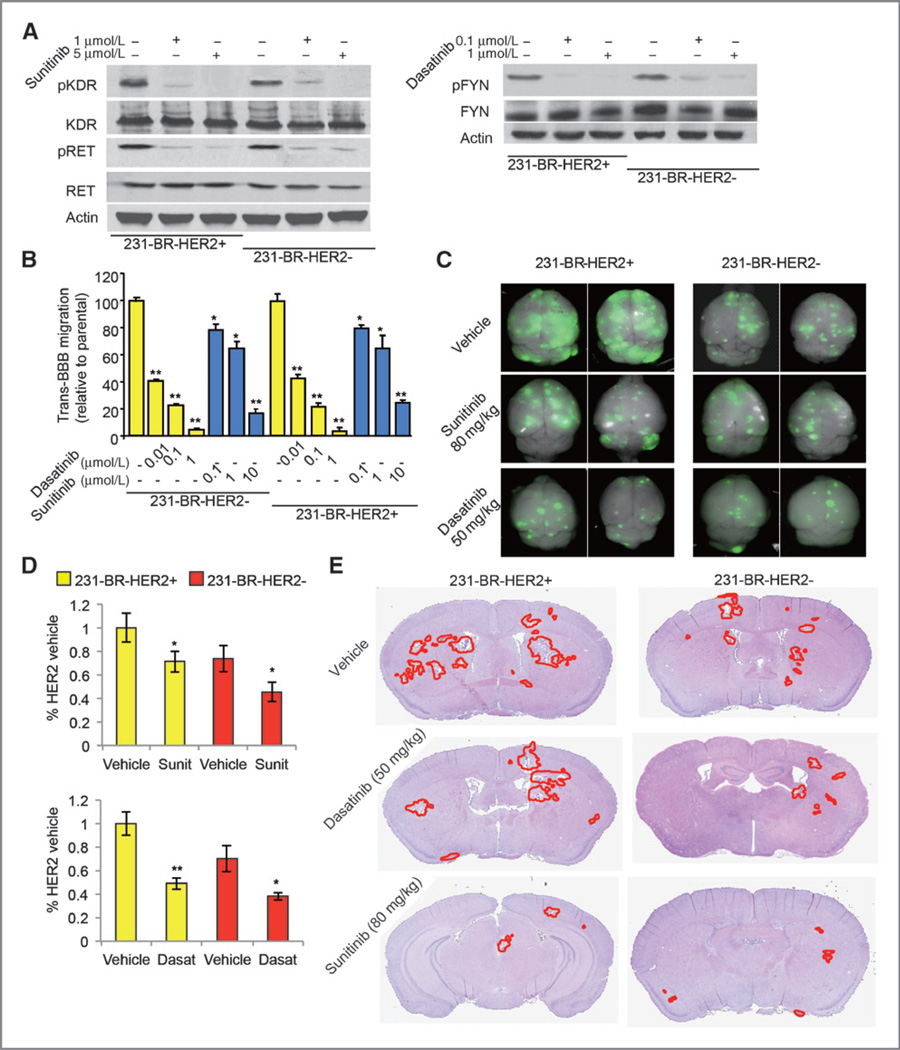
Figure 4.
Effects of sunitinib and dasatinib treatment on brain metastases. A, target inhibition by sunitinib and dasatinib on the indicated cell lines and conditions. B, in vitro BBB transmigration activity of the indicated cell lines and conditions. The number of transmigrated cells relative to the parental cell lines is plotted with n = 12. *, P < 0.05; **, P < 0.01 versus vehicle. P values were determined by one-way ANOVA. C, sunitinib and dasatinib inhibit brain metastatic colonization of 231-BR cells examined by ex vivo whole-brain imaging. The 231-BR-HER2+ or 231-BR-HER2− cells, with a retrovirus transduction expressing EGFP, were injected into the left ventricle of BALB/c nude mice. Three days after injection, sunitinib, dasatinib, or vehicle was administered by once-daily oral gavage for 28 days. Brains dissected at necropsy were imaged using a Maestro 420 Spectral Imaging System to detect the presence of EGFP-expressing metastases derived from the injected 231-BR cells. Representative dorsal whole-brain images from two mice in each treatment group are shown. D, sunitinib and dasatinib inhibit brain metastases in two 231-BR models examined by EGFP Western blot on whole-brain lysates of the animals. *, P < 0.05 versus vehicle; **, P < 0.01 versus vehicle. E, representative H&E staining images of the whole-brain sections to show the inhibition on brain metastatic loci by sunitinib and dasatinib treatment.

