. 2013 Nov 4;109(1):51–60. doi: 10.1590/0074-0276130216

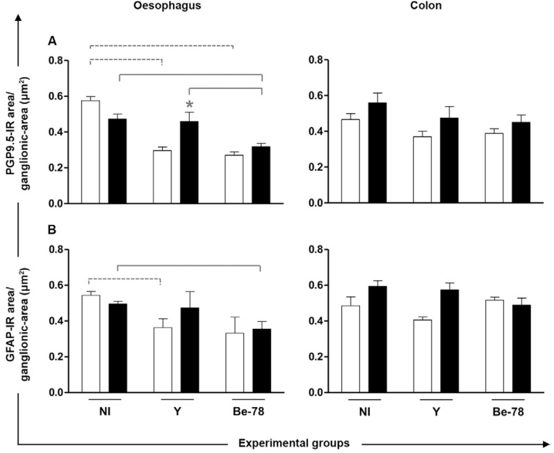
Fig. 3. morphometric analysis of Auerbach’s plexus PGP9.5-immunoreactive (IR) neurons and nerve fibres (A) and glial fibrillary acidic protein (GFAP)-IR enteroglial components (B) in the oesophagus and colon of Beagle dogs necropsied in acute (□) or chronic (■) phases from experimental infection with Y or Berenice-78 (Be-78) strains of Trypanosoma cruzi . Data are expressed as means ± standard error of the means. The dashed lines indicate significant differences between groups during the acute phase, the solid lines indicate significant differences between groups during the chronic phase and the asterisk means significant difference between acute and chronic. Groups with n = 4. Be-78: infected animals with be-78 strain; NI: noninfected animals; Y: infected animals with Y strain.

Fig. 3