. 2013 Nov 4;109(1):51–60. doi: 10.1590/0074-0276130216

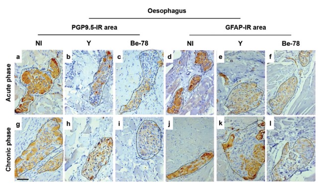
Fig. 4. photomicrographs of histological sections of the oesophagus showing Auerbach’s plexus PGP9.5-IR neurons and nerve fibres and glial fibrillary acidic protein-immunoreactive (GFAP-IR) enteroglial components in noninfected (NI) Beagle dogs or dogs infected with Y or Berenice-78 (Be-78) of Trypanosoma cruzi and necropsied in acute or chronic phases of Chagas disease. PGP9.5-IR myenteric ganglia with normal appearance in NI animals (a) and moderate reduction of IR area in animals infected with Y (b) and Be-78 strains (c) in the acute phase. Myenteric ganglia with a slight reduction in the PGP9.5-IR area in NI animals (g) or infected with the Y strain (h) and intense reduction in immunomarked area in animals infected with Be-78 strain (i) in the chronic phase. GFAP-IR myenteric ganglia with normal appearance in NI (d) and infected animals with Be-78 strain (f) and reduction in GFAP-IR area in infected animals with Y strain (e) in the acute phase. Normal GFAP-IR myenteric ganglia in NI (j) and infected animals with the Y strain (k) and reduction in GFAP-IR area in infected animals with the Be-78 strain (l) in the chronic phase. Immunohistochemistry anti-PGP9.5 in a-c, g-i. Immunohistochemistry anti-GFAP in d-f, j-l. Bar = 50 µm.

Fig. 4