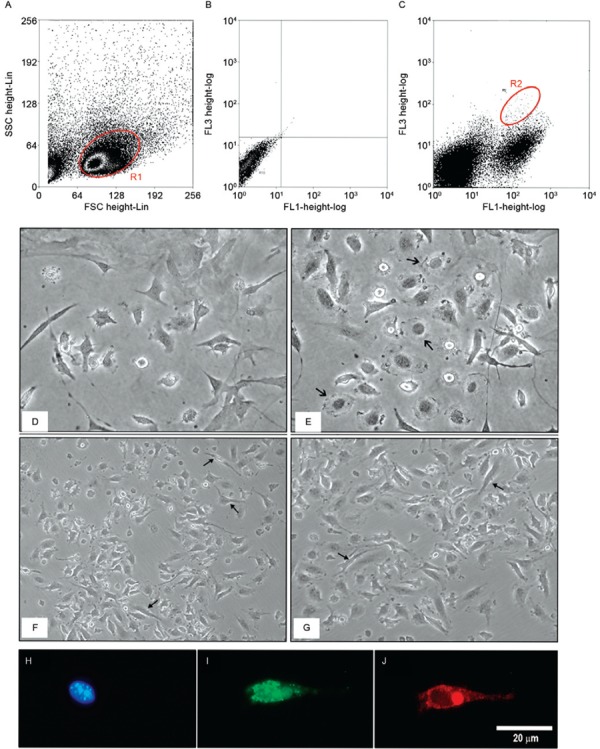
Fig. 1. : an evaluation of fibrocytes in the peripheral blood of BALB/c mice. A: dot plot [forward scatter (FSC) and side scatter (SSC)] showing retrograde (R1) marking of heat shock protein (HSP)47 + /CD45 + ; B: dot plot of the negative control in FL1/FL3; C: Dot plot identification of fibrocytes (CD45 + /HSP47 + ) in the double-positive region (R2) for FL1 {fluorescein isothiocyanate [(FITC)-CD45] and FL3 (HSP47-SPRD)}; D, E: Fibrocyte primary cultures were analysed after 21 days with phase-contrast light microscopy. Elongated cells with cytoplasmic projections and a few rounded cells (arrow) are shown at 40X magnification; F, G: fibrocyte cultures with large numbers of elongated cells with the cytoplasmic projections that are characteristic of this cell type (arrow) at 10X magnification; H: 4’,6-diamidino-2-phenylindole-stained nucleus in blue; I: fibrocytes identified by the immunostaining of CD45 (FITC) at the cell surface in green; J: HSP47 (tetramethylrhodamine) in red distributed along the cytoplasm.