
Figure 7.
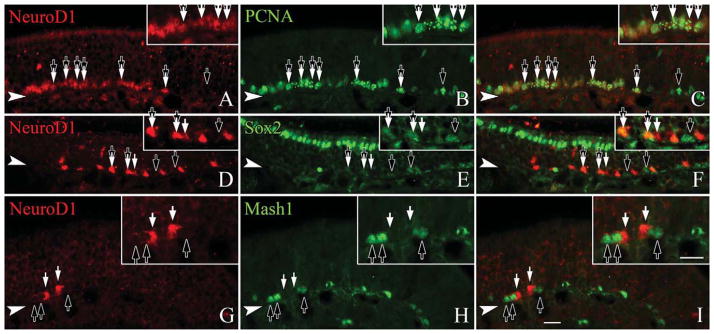
NeuroD1 is expressed in GBCs that retain cell cycle markers and lie downstream of Mash1. Sections from adult, wildtype mice are stained with anti-NeuroD1 and anti-PCNA, a marker of cell cycle progression (A–C), SOX2 (D–F), or MASH1 (G–I), as markers of GBCs. A–C: Nearly all NEUROD1-labeled cells coexpress PCNA (tandem black and white arrows), although not all PCNA-labeled cells are NEU-ROD1-positive (black arrows). D–F: Roughly one-third of the NeuroD1 labeled cells coexpress SOX2 (tandem black and white arrows), a marker for multiple subsets of GBCs, although some NEUROD1-labeled cells lack SOX2 (black arrows). G–I: NEUROD1-labeled cells (white arrows) are not labeled by anti-MASH1 (black arrows), a marker for transit amplifying GBCs. The filled arrowhead marks the basal lamina in all figures. A magenta-green version of this figure is available online as Supporting Information Figure 4. Scale bar = 20 μm.