
Figure 2.
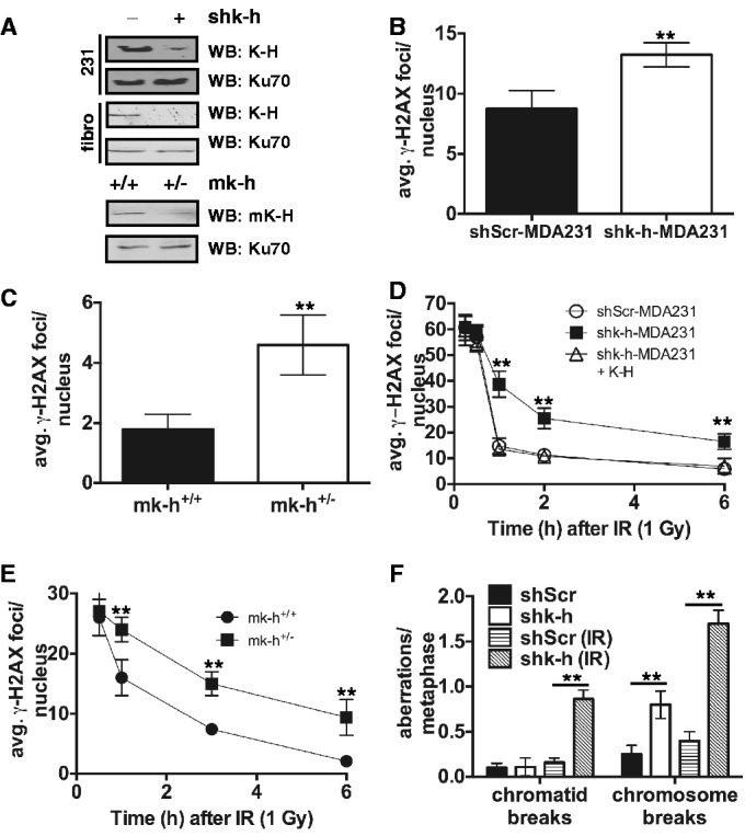
Loss of K-H leads to increased DSBs and genomic instability. (A) shScr, shk-h, shScr-MDA231, shk-h-MDA231, mk-h+/+ and mk-h+/– cells were used to interrogate the degree of K-H loss each cell system. (B and C) Basal levels of the DNA damage indicator γ-H2AX were measured in shScr-MDA231, shk-h-MDA231, mk-h+/+ and mk-h+/– cells by immunofluorescence (IF). (D and E) Rates of γ-H2AX disappearance were measured in shScr-MDA231, shk-h-MDA231, shk-h-MDA231 + K-H, mk-h+/+ and mk-h+/– cells at indicated times after ionizing radiation (IR) exposure by IF, the first time point is 0.5 h after IR exposure. (F) Amounts of genomic aberrations were quantitated in shScr and shk-h cells with and without exposure to IR (2 Gy). (**P < 0.01).