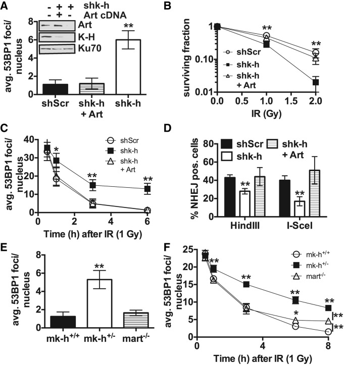
Figure 5.
Artemis is required for the DNA-repair capability in K-H deficient cells. (A) Basal levels of the DNA-damage repair biomarker 53BP1 were measured in shScr, shk-h and shk-h cells stably over-expressing Artemis (Art), (shk-h + Art), by immunofluorescence (IF). (B) Sensitivity to IR in shScr, shk-h and shk-h + Art, mock untreated or IR exposed cells was monitored by colony forming assay. (C) Disappearance of 53BP1 foci was measured in shScr, shk-h and shk-h + Art at indicated times after IR, first time point measured is 0.5 h after IR exposure. (D) The ability to perform NHEJ in shScr, shk-h and shk-h + Art was monitored by a plasmid-based NHEJ assay with plasmids digested with either HindIII to study compatible end re-ligation, or I-SceI to study incompatible end re-ligation. (E) Basal levels of 53BP1 foci were measured in wild type (mk-h+/+), K-H heterozygote (mk-h+/–) and Artemis deficient (mart–/–) MEFs by IF. (F) Rate of 53BP1 foci disappearance after IR at times indicated was measured in mk-h+/+, mk-h+/– and mart–/– MEFs by IF, first time point is 0.5 h after IR. Cells (300) were visualized for 53BP1 foci. Colonies were determined as ≥50 normal-appearing cells in a 7-day period. Events (10 000) were counted by flow cytometry. (**P < 0.01, *P < 0.05).

