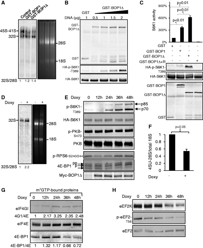
Figure 1.
Expressing BOP1Δ elicits activation of S6K1. (A) Northern blot hybridization of total RNA. HEK293 cells were transiently transfected with 1 µg of GST-BOP1, GST-BOP1Δ or GST vectors. After 48 h, total RNA was extracted and subjected to northern blot analysis using a probe specific for the precursors corresponding to the 5′-end of ITS2. The numbers below the lanes refer to the quantification for the 32S normalized to the signal for 28S (control value set at 1). (B) HEK293 cells were transfected with 0.5 µg of HA-S6K1 DNA and series of concentrations of GST-BOP1Δ DNA. After 48 h, cells were harvested, and 20 µg of lysate was analysed by western blots using the indicated antisera. (C) HEK293 cells were transfected with 0.5 µg of HA-S6K1 DNA and 1 µg of GST-BOP1, GST-BOP1Δ or GST DNA as indicated; 35 h later, where indicated, cells were treated with rapamycin (100 nM) for 45 min. HA-S6K1 was immunoprecipitated from 1 mg of total lysate. Immunoprecipitates were aliquoted into three equal portions: two were analysed by western blot to verify the IP efficiency, while the third was used for S6K1 assay. Total lysates were analysed by western blot to confirm expression of GST-tagged proteins. (D) Expression of BOP1Δ was induced by treating T-REx cells for 15 h with 1 µg/ml doxycycline. Total RNA was extracted and subjected to northern blot to detect the pre-rRNA (using the ITS2 probes).The numbers below the lanes refer to the quantification for 32S: 28S as in panel (A). (E) T-REx cells were treated with doxycycline for the indicated times, harvested and 20 µg of lysate was analysed by western blots; p85 and p70 denote the different isoforms of S6K1, which are observed here. (F) T-REx cells were treated with 1 µg/ml doxycycline for 15 h before addition of 4-SU. Total RNA was extracted after 4 h and processed to measure levels of labelled 28S rRNA, relative to total 18S rRNA, as described in the ‘Materials and Methods’ section. (G) Cell lysates were subjected to affinity chromatography on m7GTP–Sepharose and the bound material was analysed by western blot. Numbers show the quantification for eIF4G:eIF4E and 4E-BP1:eIF4E binding from a typical experiment (untreated cells value set at 1). (H) Cells were treated with doxycycline as in panels E and G. Twenty micrograms of lysate was analysed by western blot. Significance was determined by t-test.

