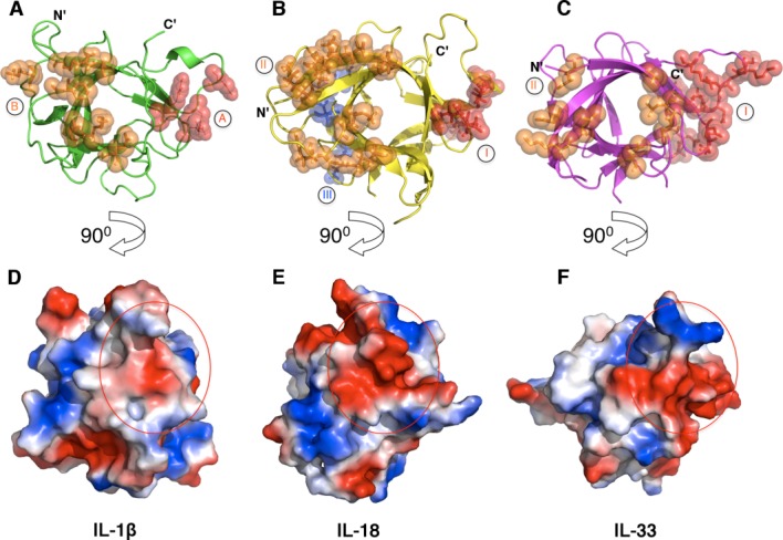
Figure 3.

Receptor binding sites on IL-1β, IL-18, and IL-33. Depicted are the secondary structures (top) and the electropotential surfaces (bottom) of IL-1β (A and D, PDB ID 1ITB), IL-18 (B and E, PDB ID 4EKX) and IL-33 (C and F, PDB ID 4KC3). Residues that have been shown to interact with their respective receptors are shown as spheres and colored in red and orange for site A (site I for IL-18 and IL-33) and site B (site II for IL-18 and IL-33), respectively. A third putative receptor-binding site (site III) on IL-18 is shown as blue spheres. The surface area of the binding site A (site I) is indicated as a red circle on each cytokine (bottom).
