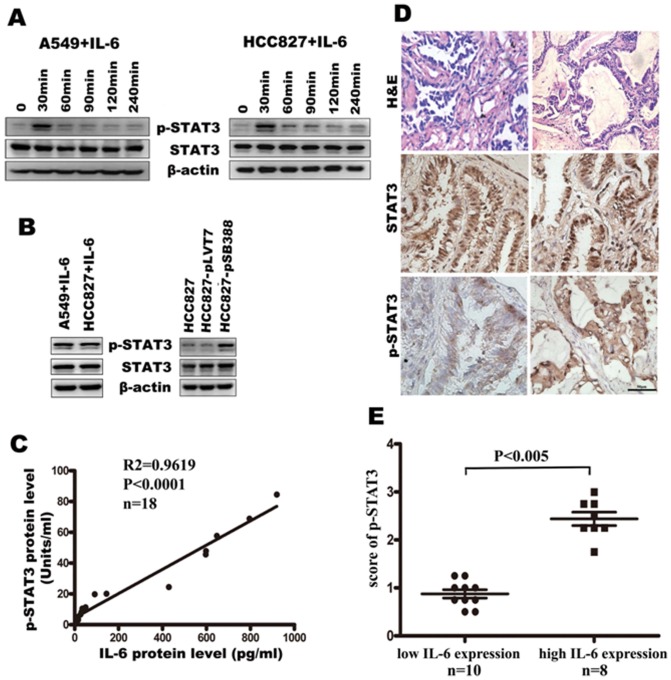
Figure 3. IL-6 induces STAT3 phosphorylation in A549 and HCC827 cells in vitro and IL-6 expression is positively correlated with STAT3 phosphorylation in patient tissure samples.
(A) STAT3 phosphorylation status in A549 and HCC827 cells after stimulation with IL-6 in a short time. (B) STAT3 phosphorylation status was determined by western blotting. A549 and HCC827 cells were stimulated with IL-6 in a long time (left). HCC827-pSB388 cells were stimulated with IL-6. HCC827 and HCC827-pLVT7 cells were used as a control (right). (C) Correlation between IL-6 production and STAT3 phosphorylation in human lung adenocarcinoma tissues (n = 18, p<0.0001). (D) Representative immunohistochemical staining for STAT3 phosphorylation in lung adenocarcinoma tissues with low IL-6 or high IL-6 expression (400 ×). (E) Comparison of the score of p-STAT3 with different IL-6 expression level.

